
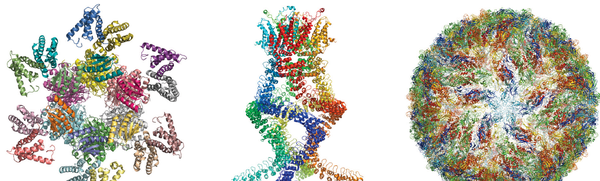
Just before noon today, Stockholm time, three real visionaries struck scientific gold: the 2017 Nobel Prize in Chemistry. The researchers had developed ways of imaging complex proteins at the atomic level, adopting electron microscopes to see how the molecules create antibiotic resistance, convert light into energy for photosynthesis and how the Zika virus functions. “For developing cryo-electron microscopy for the high-resolution structure determination of biomolecules in solution,” the Royal Swedish Academy of Sciences awarded the Chemistry Prize to Jacques Dubochet of the University of Lausanne in Switzerland, Joachim Frank of Columbia University in New York City, and Richard Henderson of the MRC Laboratory of Molecular Biology in Cambridge, England.
"This discovery is like the Google Earth for molecules in that it takes us down to the fine detail of atoms within proteins,” says chemist Allison Campbell, president of the American Chemical Society, who has done research in biomaterials. And although the Nobel Committee emphasized the biological and medical applications, Campbell says the method can be used to analyze any type of polymer, such as industrial enzymes that break down plastics.
Frank, in a phone call during the Nobel announcement, noted the practical uses were not here yet. “This is not an immediate bedside application. Several years will go by,” he said. And Campbell agrees. “This is technology on the front end,” she says. “But the potential is huge. You could understand any target molecule that you are going after. You can see its shape, and when proteins change shape they change function.”
On supporting science journalism
If you're enjoying this article, consider supporting our award-winning journalism by subscribing. By purchasing a subscription you are helping to ensure the future of impactful stories about the discoveries and ideas shaping our world today.
This is the second chemistry Nobel for microscopy in the past four years. In 2014 Stefan Hell, Eric Betzig, and William Moerner won for increasing the power of light microscopy and allowing scientists to see molecules in action within a living cell, although not at the level of atomic change.
X-ray crystallography had long been the go-to method for chemists and biologists seeking to understand the structure of proteins. It has helped scientists win more than a dozen Nobel Prizes, including the 1962 award for revealing DNA’s double helix, according to a news article in Nature, and by 2015 x-rays had been used to determine the structure of about 90 percent of the approximately 100,000 molecules in the popular Protein Data Bank.
But the technique cannot do everything. As its name implies, crystallography requires its targets to be made into crystals. And with many large, complicated molecules found in and around cells—uch as ribosomes, which turn genetic instructions into working proteins—scientists simply could not make that happen.
Electrons, however, can bounce off every atom in a protein and reveal its structure. That structure is three dimensional, and beginning in the 1970s Frank developed a mathematical image-processing method that allowed a computer to merge several two-dimensional electron microscope images into a sharp 3-D picture. Dubochet’s contribution was to show how this kind of microscopy could be used on biomolecules. Molecules such as proteins are surrounded by water that helps them maintain their structures, but electron microscopy dried up the water. Dubochet figured out a way to cool the water rapidly so it became like glass—the form is called vitrified water—and allowed the molecules within to retain their shapes.
Then in 1990, after 15 years’ work refining sample preparation and electron detection, Henderson succeeded in using an electron microscope to create an image of a large bacterial cell membrane protein called bacteriorhodopsin, and do it at atomic resolution. Henderson, with Nigel Unwin, wrote in Scientific American’s February 1984 issue about how the two pioneered the use of electron microscopes to see the details of cell membrane proteins.
Now for any scientist “who is interested in a protein’s structure and function—well, I’d love to have one of these in my laboratory,” Campbell says. Dubochet, Frank, and Henderson will get their medals and each a third of the 9 million Swedish krona (about $1.1 million) prize at the annual Nobel ceremony in Stockholm in December.