
In 1231 Frederick II, the Holy Roman Emperor who ruled over much of Europe, issued a decree requiring schools that trained doctors to hold a human body dissection once every five years. It was a slow debut for what would become a cornerstone of medical education. During the Renaissance, cadaver dissections helped scientists and artists gain a hands-on understanding of human anatomy. Today they are an essential experience for first-year medical students, a time-honored initiation into the secrets of our flesh.
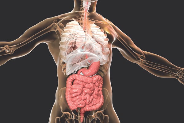
Now, nearly a millennium after its measured introduction, cadaver dissection may have begun an equally slow exit. This year a few U.S. medical schools will offer their anatomy curriculum without any cadavers. Instead their students will probe the human body using three-dimensional renderings in virtual reality, combined with physical replicas of the organs and real patient medical images such as ultrasound and CT scans.
The program developers hope technology can improve on some of the limitations of traditional approaches. It takes a long time to dissect cadavers, and some body parts are so inaccessible that they may be destroyed in the process. Plus, the textures and colors of an embalmed cadaver's organs do not match those of a living body, and donated bodies tend to be old and diseased. “If you want to be truthful about anatomy education, it hasn't changed much since the Renaissance,” says James Young, chief academic officer of the Cleveland Clinic Lerner College of Medicine, a program in collaboration with Case Western Reserve University that opened a new cadaverless campus this summer. “But as technology advanced and as knowledge increased, there came a push to do things better and faster and give students a more appropriate representation of human anatomy.”
On supporting science journalism
If you're enjoying this article, consider supporting our award-winning journalism by subscribing. By purchasing a subscription you are helping to ensure the future of impactful stories about the discoveries and ideas shaping our world today.
Young, who studied medicine in the 1970s, experienced a “massive disconnect” between his own anatomy education and what he saw during clinical training in cardiology. When he tried to access organs in living patients, looking at imaging results or footage from tiny inserted cameras, he found the inside of human bodies did not match what he had seen in cadavers. “They're totally different,” Young says. “The embalmed cadaver has a very flat, compressed organ presentation. The colors are not the vibrant colors of a living human.” The difference can distract from learning, he says.
Virtual anatomy tools, in contrast, provide a more faithful view of living organs, helping students form a foundational understanding of the body's structures, Young and other medical educators say. By donning VR headsets or augmented-reality goggles, which show digital imagery superimposed on the real world, students can examine an organ from all angles. They can connect structure with function by watching a beating heart or moving joints. They can also select views that add other organs or the entire circulatory and nervous systems to better see relations among structures. “I was amazed,” says Mark Schuster, dean of Kaiser Permanente School of Medicine in Pasadena, Calif., which will welcome its first class of medical students in 2020. “I wished I had that when I'd been learning anatomy. It really helped make it all come together.” His program's first-year students will have a cadaverless curriculum.
Adopting high-tech alternatives makes sense for brand-new medical programs that have neither the tradition nor the facilities for cadaver dissection, but even some existing ones are adopting digital tools to supplement their anatomy courses. “The big advantage I see is that the visuals are very clean,” says Darren Hoffman, an assistant professor of anatomy and cell biology, who uses interactive 3-D anatomy software in his courses at the University of Iowa Carver College of Medicine. “That helps building your 3-D mind's eye of the body, so that when you look at a patient's ankle, you know what's underneath the surface and how it's all related.”
Besides the educational advantages, going cadaverless is an economic decision for new programs. It costs several million dollars to build a cadaver laboratory, which requires a lot of space, as well as safety measures that meet legal regulations. And although cadavers are donated, medical schools still pay for preparation, maintenance and, eventually, burial. These costs are an even bigger challenge for schools in less wealthy nations, Young says. What is more, many countries still face a shortage of donations and rely on unclaimed bodies for dissection, according to a 2018 study.
Cadaverless anatomy education has its drawbacks. It may be hard to develop a perception of depth in a virtual body, and students will miss out on seeing bodies' natural anatomical variations, according to Hoffman. Students may also lose the emotional, even philosophical impact of working with a cadaver, commonly seen as a doctor's first patient. “There's a sort of awe and respect that comes from that,” Hoffman says. “You recognize how amazingly cool and intricate the human body is, and you start to realize that everybody on the planet is this amazing—and so am I.” Yet the lab is not the only way to forge a student's medical identity, Hoffman and others say. For instance, students could interact with living patients earlier in their studies.
Another open question is whether students learn as well using the digital tools. Educators' studies are probing whether replacing old techniques with new technology will actually improve, and not harm, the quality of their students' education. Their results, if positive, may encourage more schools to convert. “It feels early to call this a trend, but given the sheer number of schools that have shown interest, it feels like something's happening,” Schuster says.
Anatomy education has been resistant to change for so long that Young and others see what is happening now as a sign of a possible historic transition. “We're at the beginning of a paradigm shift, no question about that,” Young says. “That shift is going to take several years. But if you asked me how is anatomy education going to be done in a decade? It's not going to be done with cadavers. That's my prediction.”
