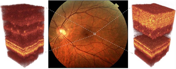
Sightseeing: Adaptive optics could peer deep into damaged eyes for earlier diagnoses
The earlier diabetic retinopathy, macular degeneration and glaucoma—the three most prevalent diseases causing blindness—can be diagnosed, the more successfully they can be treated.
PHOTOS BY ROBERT J ZAWADZKI, UC DAVIS
Join Our Community of Science Lovers!
On supporting science journalism
If you're enjoying this article, consider supporting our award-winning journalism by subscribing. By purchasing a subscription you are helping to ensure the future of impactful stories about the discoveries and ideas shaping our world today.
The earlier diabetic retinopathy, macular degeneration and glaucoma—the three most prevalent diseases causing blindness—can be diagnosed, the more successfully they can be treated. Obtaining high-res images of the retina is not so easy, however, due to aberrations caused by imperfections in the cornea and crystalline lens.
"There is a whole history of attempts to image the retina in a way that would help doctors diagnose blinding diseases earlier," says physicist Scot Olivier, who is leading the LLNL work. OCT can make noninvasive, in vivo measurements of the thickness of specific retinal layers, such as the nerve fiber layer, which thins in patients with glaucoma.
Most ophthalmologists already use OCT to measure nerve fiber thickness, Olivier says. If a company could commercialize the adaptive optics as an add-on to OCT, imagine a new generation of devices allowing volumetric retinal imaging with high sensitivity.
Subscribe to Support Independent Journalism
Great science journalism requires human expertise, time, effort and creativity. And it costs money. That’s why I and the journalists here at Scientific American hope you’ll join our community.
When you subscribe, you are supporting staff and freelance journalists who are passionate about telling science stories that are true, important and compelling. Our editors and reporters are often experts in their fields, which means they understand the nuances of big discoveries and can untangle the breakthroughs from the hype. With a subscription, you are also supporting rigorous fact-checking to ensure the words we publish are precise and accurate. And you’re supporting original illustrations, graphics and photos that bring you closer to an advanced laboratory, an ice sheet in Antarctica or a space mission in orbit. You’re helping us craft other types of high-quality journalism as well: Our newsletters are carefully written, edited and curated by staffers you have or will come to know and love. Our Science Quickly podcast is based on original reporting, collaboration with editors and scientists and exacting production.
Subscriptions keep this engine running so we can continue to deliver thoughtful, rigorous and independent science journalism to you. In an era of viral misinformation, this work is crucial. If you value what we do, I hope you’ll consider joining us as a subscriber
Thank you,
Jeanna Bryner, Editor in Chief, Scientific American