Due to its transparent nature, a zebra fish embryo in its fourth day of gestation can provide scientific data on the exact time when certain facial malformations, such as cleft lip, occur. This image, which took first place this year, was taken with a confocal microscope using 10x magnification. Credits: Oscar Ruiz; University of Texas M. D. Anderson Cancer Center
Small Wonders: Science Meets Art Under the Microscope [Slide Show]
Share
ZEBRA FISH EMBRYO Due to its transparent nature, a zebra fish embryo in its fourth day of gestation can provide scientific data on the exact time when certain facial malformations, such as cleft lip, occur... Oscar Ruiz; University of Texas MD Anderson Cancer Center
AGATE Retired biologist Douglas Moore won second place for photographing the delicate details recorded on a 270 million-year-old agate from Canyon Tepee, S.D. These stones are unique not only for their age and vibrant colors, but because they often contain fossil remains that other types of agate don’t have... Douglas L. Moore; University of Wisconsin, Stevens Point Museum of Natural History
NEURONS Rebecca Nutbrown from the University of Oxford took third place with this image of neurons (green) derived from human skin cells. The image also shows Schwann cells (in purple), which give support to the neurons, interacting in the same way they would with neurons in the brain... Rebecca Nutbrown; Nuffield Department of Clinical Neurosciences, University of Oxford
PROBOSCIS OF A BUTTERFLY This photo shows the proboscis of a butterfly, taken with an image-stacking technique. Jochen Schroeder; Chiang Mai, Thailand
Advertisement
THE LEG OF A WATER BEETLE This colorful photo shows the front side of the leg of a water beetle. The image was produced with a confocal microscope, using a technique in which a scanner is used to photograph sections up to 250 nanometers thick... Igor Siwanowicz; Howard Hughes Medical Institute (HHMI)
BUBBLES OF ASCORBIC ACID This image shows bubbles formed by melted crystals of ascorbic acid (vitamin C). The photo was made using a technique involving polarized light. Marek Mis; Marek Mis Photography, Poland
"ROSE OF JERICHO" Leaves of Selaginella , a genus of plants common in tropical areas, are pictured here. One species, S. lepidophylla , is commonly known in Latin America as “rose of Jericho.” The image was taken using a technique called differential interference contrast to visualize transparent structures... David Maitland; Norfolk, United Kingdom
WILDFLOWER STAMENS Israeli photographer Samuel Silberman captured this colorful image of wildflower stamens, the male reproductive organs of a flower. The photo used fiber-optic lighting, in with a light source brought to the subject through flexible tubes... Samuel Silberman; Yahud–Monoson, Israel
Advertisement
COFFEE This image of coffee crystals was made with a technique in which light passes under a microscope through a polarizing filter. Vin Kitayama and Sanae Kitayama; Vinsanchi Art Museum Azumino, Nagano, Japan
UNICELLULAR ORGANISM This image shows a unicellular organism belonging to the genus Frontonia , common in freshwater and saltwater environments around the world. It was taken with a technique called differential interference contrast, with a 200x magnification... Rogelio Moreno Gill; Panama
Centuries after its invention, the microscope continues to prove that it is not only crucial to science but can also produce works of art—a feat acknowledged each year by the Nikon Small World Award, a competition in which the famous photographic brand recognizes what judges consider the most interesting and beautiful scientific images.
Microscopic pictures of an insect, a 273 million-year-old rock and a culture of human neurons are among the 2016 winners. “Each of these images evoked a powerful reaction of wonder, tenderness or discomfort in the judges. This is where the connection between art and science, the emotional and the technical lies,” says Eric Flem, communications manager of Nikon Instruments.
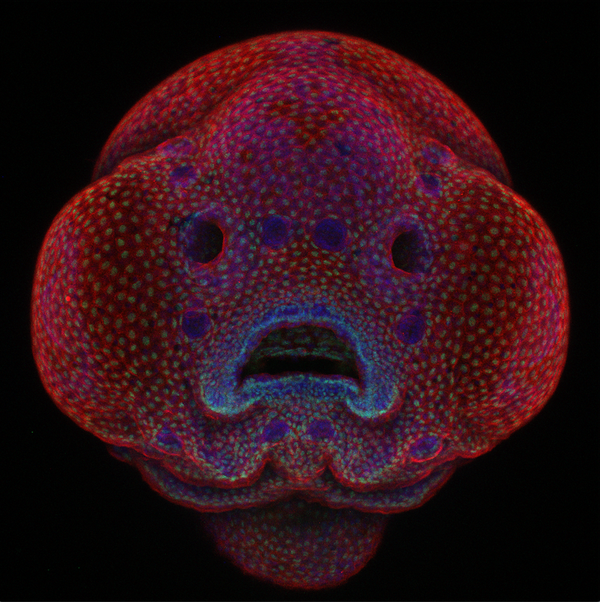
Oscar Ruiz, an Mexican-American scientist at the University of Texas M. D. Anderson Cancer Center in Houston, was the first-place winner this year with a photograph showing a zebra fish embryo at four days of gestation. Ruiz, who specializes in the genetic mutations that cause cleft lip and palate, is working on a study to explain how these physical abnormalities develop during pregnancy. To explore this issue the researcher uses zebra fish, whose transparent embryos offer almost perfect visualization of internal processes. His winning image involved one of these subjects, and there is more to it than visual beauty. The observational techniques Ruiz created make it possible to see how an embryo changes in real time. “It is something with high scientific value because it has not been done before with the cleft lip,” he says.
Ruiz was thrilled to take the top spot in in the photo competition—organizers say judges reviewed 2,000 images from 70 countries. “This motivates me to keep working even more. Images like this help us to find the exact moment when the problem occurs, and then provide the basis for a solution,” Ruiz says.
According to tradition, the images will be compiled in a calendar that is distributed among laboratories around the world. “I collected this calendar for years,” Ruiz quips. “Knowing that my image will now be there is priceless.”