
On supporting science journalism
If you're enjoying this article, consider supporting our award-winning journalism by subscribing. By purchasing a subscription you are helping to ensure the future of impactful stories about the discoveries and ideas shaping our world today.
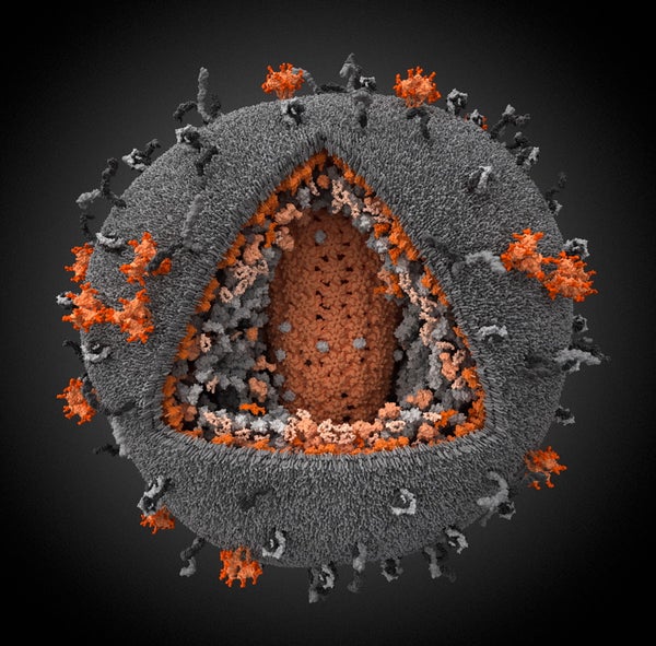
Touted as the most accurate model to date of the human immunodeficiency virus (HIV), this 3-D digital simulation is among the winners of the 2010 Science and Engineering Visualization Challenge, sponsored by Science and the National Science Foundation.
The model takes into account the latest knowledge of HIV's structure and biology collected from more than 100 scientific papers, according to its creators at the Visual Science Company in Moscow.
The color orange is used to indicate parts of the particle encoded by the virus genome, whereas gray represents parts captured from the host cells. The protruding orange structures are surface glycoprotein trimers, which allow HIV to bind and fuse with the host cell. The viral plasma membrane is mostly co-opted from that of the host cell as newly formed viruses bud from infected cells. The orange structure at the center of the model is the capsid, a cone-shaped pod that holds the viral RNA and enzymes. These enzymes include reverse transcriptase, a major target for current antiretroviral drugs.
The HIV particle has a diameter between 100 and 180 nanometers. (A nanometer is one billionth of a meter.) Its genome consists of just nine genes encoding 15 different proteins. Some 33.3 million people around the world carry the virus.
"It's looks like a fuzzy ball of yarn, but as you look at it more closely it's an incredibly intricate display of the proteins from the virus itself and from the host it infects, in a very dramatic way," said Colin Norman, news editor at Science, in an interview posted to the journal's Web site for media. The winning images are set to be published February 18 in Science.
—Nina Bai
