
Ink, watercolors and pencils may not seem like critical tools for neuroscientists. Yet these were the implements that helped Santiago Ramón y Cajal, the father of modern neuroscience, discover that neurons are the main functional unit of the brain. His intricate drawings of brain cells served as a lens through which he could examine how their structure dictated their function.
Ramón y Cajal's late 19th- and early 20th-century sketches were nothing like the bland illustrations that fill most modern textbooks. They were works of art in their own right—with delicate snaking tendrils of black ink branching out from individual cell bodies and becoming tangled with neighboring neurons. They are still heralded for their power to showcase the inherent beauty of the nervous system.
Today's illustrative tools are more technical. Scientists use chemical stains to imbue cells with a fluorescent glow or imaging machines to highlight brain activity with bursts of color. But the results are often the same: spectacular visuals that help us better understand the inner workings of our mind.
On supporting science journalism
If you're enjoying this article, consider supporting our award-winning journalism by subscribing. By purchasing a subscription you are helping to ensure the future of impactful stories about the discoveries and ideas shaping our world today.
Few images gathered in the course of research ever escape the lab, but the annual Art of Neuroscience Competition helps to bring some to light. The contest, organized by Tycho Hoogland, Chris Klink and Cathrin Canto, all at the Netherlands Institute for Neuroscience, recognizes one grand prize winner and four honorable mentions from a wide selection of submissions, all intended to join art with science in some way. What follows is a subset of this year's entries—some winners and others we felt were particularly beautiful or revealing. All the submissions are available to view at http://aon.nin.knaw.nl.
Grand Prize Winner
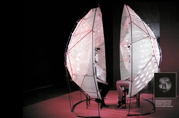
1. The Mutual Wave Machine
Click or tap to enlarge

OLEG BORODIN; COURTESY OF MATTHIAS OOSTRIK (inset)
You are enveloped in darkness. A spray of light erupts in front of you, cast onto a translucent white screen. It pulses as it coalesces into a face—your face. This spectacle is the Mutual Wave Machine's grand finale, seen only by partners who, when sitting inside the contraption together, can get literally on the same wavelength. Headsets measure each person's brain waves using tiny EEG monitors. As their frequencies align, patterns emerge in the projected light. Greater synchrony produces more vivid images, progressively morphing into images of their faces.
Researcher Suzanne Dikker of New York University and Utrecht University in the Netherlands and artist Matthias Oostrik created the installation, which has now been exhibited around the world. It makes immediate what is usually amorphous: a personal connection between two people.
Honorable Mentions
2. Axons in Shape

MICHIEL KLEINNIJENHUIS FMRIB, University of Oxford
A protein called myelin insulates axons to make them better electrical conductors. In this cross section, prepared by postdoctoral MRI physicist Michiel Kleinnijenhuis of the University of Oxford, a cluster of individual axons appears red, with sheathes of white myelin wrapped around them. The view makes it easier to see their variable size and shape. The diameter and density of an axon, among other properties, determine how well it works and are useful measures in studying brain functionality.
3. Exploding Brain Myths

COLLABORATION BY RHIANNON MEREDITH, RHODÉ VAN WESTEN AND MATTHIJS VERHAGE AND ANIMATORS JON HUNTER, MATT PARTRIDGE, HENRY PAKER AND GARETH GWYNN; FUNDED BY A VU UNIVERSITY RESEARCH FELLOWSHIP AND A NETHERLANDS ORGANIZATION FOR SCIENTIFIC RESEARCH AWARD
Brains are enormously complex, and myths about them abound: We only use 10 percent of our brain. Left-brained people are more logical, right-brained more artistic. There is one hormone that makes us fall in love. And so on. Neuroscientist Rhiannon Meredith of VU University Amsterdam in the Netherlands created a video series, available via YouTube, to set the record straight. This still image comes from the final video, which explains why brain-training games do not increase your overall intelligence. Learning something new is thought to change only those cells directly involved in the activity, as shown in the illustration.
4. Aurore Boreale

Credit: ALICIA LEFEBVRE
In this painting by French artist Alicia Lefebvre, a slice of the hippocampus resembles the famous Northern Lights. This brain region has well-defined layers, each of which features particular types of cells, depicted here in different hues. These cells serve relatively distinct roles in memory consolidation—the process by which the brain converts memories from short to long term. The area is also one of the first to be damaged in Alzheimer's disease.
5. Butterfly of the Soul

ROBIN SCHARRENBERG RG Neuronal Development, Center for Molecular Neurobiology Hamburg (ZMNH), University Medical Center Hamburg-Eppendorf
Santiago Ramón y Cajal described neurons as “mysterious butterflies of the soul,” a phrase that research assistant Robin Scharrenberg says stuck in his mind as he studied mouse pyramidal cells, such as the one shown here, under the microscope at the Center for Molecular Neurobiology Hamburg at the University Medical Center Hamburg-Eppendorf in Germany. He was trying to understand how structural changes in these cells—one of the most common neurons in the neocortex—might contribute to functional changes akin to those found in autism spectrum disorder. As is clear from this image, pyramidal cells typically feature a long axon that stretches out to deliver signals to other cells and shorter, branched dendrites that stay close to the cell body to receive messages.
Editors’ Picks
Perhaps the best works of art are those that reveal some truth about their content. Scientific American Mind editors selected the following images not only for their visual appeal but also for what they reveal about the brain. They represent a range of scientific ideas and imaging techniques and sit at the very intersection of art and neuroscience.
6. Synaptic Sense of Sound

SONJA PYOTT Department of Otorhinolaryngology, University Medical Center Groningen
When we hear a sound, it enters the ear as a physical wave in the air but must become a chemical signal for our brain to understand it. Hair cells—shown here in green—perform that critical translation. These cells have tiny, fingerlike protrusions that wiggle as waves pass them by. This movement opens and closes minuscule channels in the cell, starting an electrical signal—which in turn becomes a chemical neurotransmitter signal—that travels to the brain's auditory-processing areas for interpretation. Neuroscientist Sonja Pyott of the University Medical Center Groningen in the Netherlands used fluorescent markers to highlight each structure as part of her research on preventing and reversing hearing loss.
7. Stellate Ganglion on Fire

PAMELA IMPERADORE Stazione Zoologica Anton Dohrn
Between the brain of an octopus and its peripheral nervous system sits a collection of nerves called the stellate ganglion, shown here in reddish-orange. Only two pallial nerves connect to the stellate ganglion. Severing one eliminates muscle control, but thanks to advanced regeneration abilities, the octopus can regrow nerves and regain the connection in just a few months. This image, created by Ph.D. student Pamela Imperadore of the Stazione Zoologica Anton Dohrn in Naples, Italy, shows a stellate ganglion only a few days after one nerve was severed.
8. Moments Meant to Pass

Credit: LUKE MANINOV HAMMOND
The smattering of black in this image looks like a harmless splash of paint but is actually one of the key agents behind Alzheimer's. Amyloid plaques—clumps of misfolded proteins that form in the spaces between neurons—can develop in normal brains. If they are not cleared and start to accumulate, though, they disrupt brain function. This image by Luke Maninov Hammond, microscopy facility manager at Queensland Brain Institute in Australia, came from a study conducted by Jürgen Götz and his colleagues investigating whether ultrasound could reduce amyloid plaques in mouse brains. Red and blue microglia cells, the primary type of immune cell in the brain, stand in contrast with the black plaque.
9. Brain Impressionism

SOLEDAD DE OLMOS Medical Research Institute Mercedes and Martin Ferreyra (INIMEC-CONICET–National University of Córdoba)
This black, scraggly neuron, floating in a sea of pink, resembles a dying tree—and in fact, it is degenerating. Its color comes from a stain called amino-cupric silver, which was originally used to identify neurodegenerative disorders because only dying neurons absorb it. Neuroscientist Soledad de Olmos of the Medical Research Institute Mercedes and Martin Ferreyra in Argentina made the image by infecting a mouse with a virus that causes brain inflammation, which can make neurons too excitable. This overexcitability can cause excitotoxicity, in which nerve cells fire so frequently as to damage their own structure. Excitotoxicity is involved in a number of neurodegenerative disorders such as Alzheimer's, multiple sclerosis and amyotrophic lateral sclerosis.
10. Heart Space

IMAGE BY ELIZABETH JAMESON, WITH ASSISTANCE OF DEPARTMENT OF NEUROLOGY, UNIVERSITY OF CALIFORNIA, SAN FRANCISCO
In this image, artist Elizabeth Jameson reclaims the brain scans used to diagnose and track her own case of multiple sclerosis. This particular scan is a type of MRI called diffusion tensor imaging that looks at the flow of matter in the brain. As Jameson played around with the scan, she stumbled on this image of a heart, which filled her with an instantaneous sense of wonder. She says she was uplifted by this symbol of love and compassion embedded in an analytic scan of her diseased brain.
