
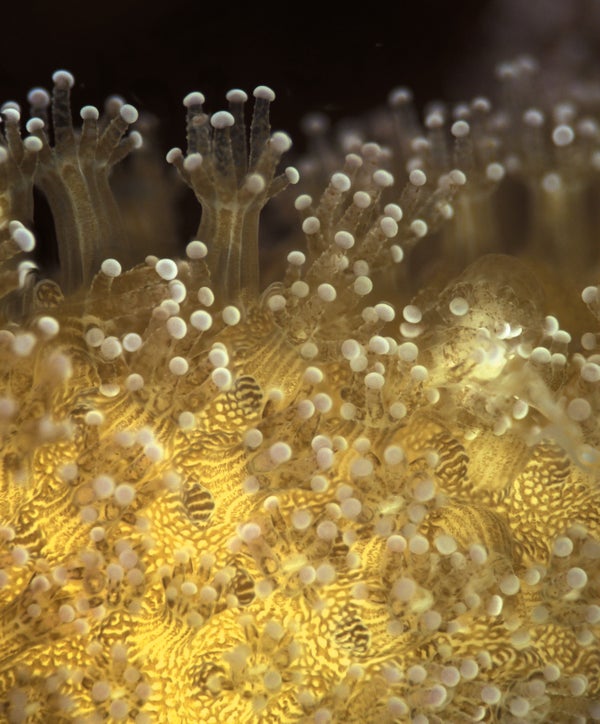
Underneath the waves of the Pacific Ocean, scientists in 2015 watched a slow and surprising dance of death in unprecedented detail.
Andrew D. Mullen, a graduate student who works with oceanographer Jules Jaffe at the University of California, San Diego's Scripps Institution of Oceanography, was peering at a dying coral reef near the island of Maui in Hawaii. The reef had just been “bleached”—symbiotic algae that live in each tiny coral polyp got too hot, and the coral had expelled them. “The polyps turned white, and they were weak. But they were still alive,” Mullen recalls.
Then doom arrived, in the form of green threads of other, invasive algae that drifted onto ridges of calcium compounds between the polyps. “Even weak coral polyps should be able to resist algae,” Mullen says. But researchers had never seen how, in the wild, algae settle the in-between spaces and over months expand to cover the polyps. “Once they are overgrown, corals don't stand a chance,” he explains. “They are covered in a green film, and at that point, they're dead.”
On supporting science journalism
If you're enjoying this article, consider supporting our award-winning journalism by subscribing. By purchasing a subscription you are helping to ensure the future of impactful stories about the discoveries and ideas shaping our world today.
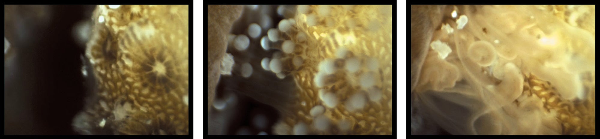
Microscope images show Stylophora (right side of pictures) being eaten by a rival coral, Platygyra (left side), which has extruded its curtainlike stomach to dine.
This life-and-death waltz occurs on a minuscule stage. Each polyp is about one millimeter wide. The invading algae filaments can be a twentieth of a millimeter—similar to the width of a human hair—and the symbiotic algae expelled by the corals were 10 times smaller. Mullen and Jaffe were able to see all of them using a new kind of magnifier in a waterproof housing: the Benthic Underwater Microscope, the first instrument powerful enough to get real-time views of these creatures at the bottom of the sea.
Jaffe and other ocean scientists had been able to see coral polyps close up before but only in the laboratory. On the seafloor, conditions are too dark and difficult for ordinary microscopes to produce clear images. Mullen's solution was to mount several bright LED lights around the device and to use an electrically adjustable focusing lens with a flexible polymer that rapidly flattens or thickens to keep a tiny object in sharp view, like the lens of a real eye.
The lens has shown not just death but also strategies for life. Jaffe and Mullen have watched battles between competing corals in which one digests its neighbor (above). This does not occur, however, when the corals are of the same species, indicating the animals somehow sense their kin. How this happens remains to be seen, but with newly opened eyes, the scientists are certainly going to look.
Underwater Microscopy
The seafloor is anathema to microscopy. Down there, dark waters and shifting currents conspire to make any minuscule object a blurry smear of motion with most lenses. The Benthic Underwater Microscope’s unique design solves these problems. Its centerpiece is a deformable lens (1) that can change its shape every few milliseconds, flattening or bulging to adjust focal length and keep images sharp. To illuminate targets, the system uses submillisecond light pulses from a ring of LEDs (2) , providing rapid, minimally disruptive exposures free of motion blur. By combining exposures taken at different focal lengths, a single image can be made in which all parts of a subject are in focus (3).
Click or tap to enlarge

Credit: JOHN GRIMWADE; Source: “Underwater Microscopy for In Situ Studies of Benthic Ecosystems,” by Andrew D. Mullen et al., in Nature Communications, Vol. 7, Article No. 12093; Published online July 12, 2016.