
Miniature lab-grown brains, called brain organoids, open little windows into human neurodevelopment that are impossible to get by studying actual human brains. But without a blood supply or sensory input, these pea-sized neuron clusters eventually hit a developmental ceiling. And because the organoids lack a body, scientists can’t easily connect their brain activity to observable behavior, limiting their usefulness. But a new study shows a way to give organoids some of what they’ve been missing—by transplanting them into mice.
In a study published today in Nature, scientists report that human brain organoids, cultured from stem cells, can integrate with the mouse brain and form functional neural circuits. The result is “absolutely transformative,” says Simon Hippenmeyer, a neuroscientist at the Institute of Science and Technology Austria, who was not involved in the new study. The approach “promises many future applications for ... using human neurons to not only study development but also model neurodevelopmental diseases,” he says.
This isn’t the first time human brain cells have been transplanted into mice. But organoids, like the human brain itself, grow much slower than mouse brains. “They’re always outcompeted,” says Stanford University neuroscientist Sergiu Pașca, senior author of the new study. To get around this, he and his colleagues deleted a gene in mouse embryos that controls the formation of two major brain regions, the cerebral cortex and the hippocampus. These animals were born with only about half their normal brain volume.
On supporting science journalism
If you're enjoying this article, consider supporting our award-winning journalism by subscribing. By purchasing a subscription you are helping to ensure the future of impactful stories about the discoveries and ideas shaping our world today.
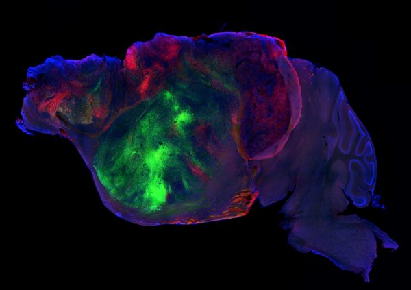
Into each mouse’s empty intracranial space, the researchers placed four organoids, each of which contained about 100,000 neurons. Within a few months, these had divided into roughly four million neurons, and the cells had sent axons—fibers that convey electrical signals—throughout the mouse nervous system all the way to the spinal column. It’s unclear how much the human neurons interacted with their mouse counterparts, but brain imaging showed patterns of coordinated activity across the human graft. “This tells us that the cells are active and they’re communicating with each other,” Pașca says.
Most strikingly, the organoids produced von Economo neurons, a class of brain cells thought to be important for social cognition in primates, elephants and dolphins. These cells have never been documented in organoids confined to a petri dish, suggesting that the mouse-brain environment enables human neurons to develop more fully.
Alysson Muotri, a geneticist at the University of California, San Diego, who was not involved in the new study, says it represents a “tour de force in terms of technology.” But he argues that it’s hard to disentangle the human neurons’ activity from that of the rodent neurons, which could complicate efforts to probe human brain function and malfunction. “I would rather bet on a fully humanized model system instead of a chimera to study human diseases,” he says, noting that, as organoids improve, it may be possible to “‘teach’ circuits to learn and memorize actions that might replace complex behaviors observed in mice.”
Still, the transplants open new possibilities for investigating neurological conditions, from autism to dementia, that conventional models don’t provide, Paşca says. “We built a system so that it captures aspects of disease that would be difficult to capture with any of the other systems,” he says.