
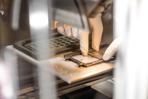
Knowing how proteins behave inside individual human cells can tell us whether those cells will live, die or malfunction—information that can serve as an early warning of disease. But such detailed information is hard to come by because today's analytical methods require minimum samples of hundreds to thousands of cells. Now, however, researchers at Pacific Northwest National Laboratory (PNNL) have developed a “molecular microscope” for detecting and identifying proteins within samples of only a few cells—or even a single cell—and have used it to distinguish between diseased and healthy tissue.
The new apparatus can analyze tissue samples 500 times smaller than those needed for other protein-identification technologies. In one test, scientists used the tool to detect roughly 650 proteins in a single human lung cell. In another, they used it to inspect small samples of human pancreas tissue in a lab dish to determine if the tissue was diabetic or healthy. (Diseased pancreatic cells can block the production of insulin, causing type I diabetes.) The researchers have also employed the technology to identify thousands of proteins from small numbers of healthy human brain, lung, liver and uterus cells. They hope this approach will lead to more targeted and personalized treatments for diseases.
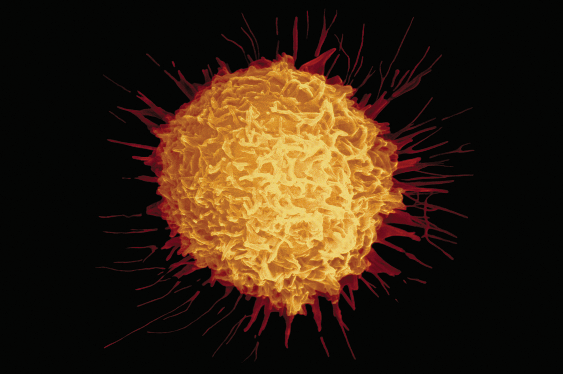
Prostatic cancer cell. Credit: Science Source
On supporting science journalism
If you're enjoying this article, consider supporting our award-winning journalism by subscribing. By purchasing a subscription you are helping to ensure the future of impactful stories about the discoveries and ideas shaping our world today.
The system first collects tissue samples in minuscule wells etched into the surface of a glass chip. Then a robotic arm dispenses droplets of chemical reagents—for extracting and isolating proteins—into each hollow. Scientists then use a device called a mass spectrometer to identify the proteins.
Until now, protein-identification technologies could provide only an overall view of larger samples rather than detailed information about the proteins in single cells or very small cell clusters. “We can now isolate and analyze the individual needles from the haystack, whereas before we had to analyze the needles and haystack together,” says chemist Ryan Kelly. He and his colleague Ying Zhu, both then at PNNL, co-authored the study published this past summer in Angewandte Chemie.
The main reason that working with samples this small is difficult is because some material is lost during each processing step. The new system addresses this issue by using wells that minimize the surface area onto which proteins might stick. David Goodlett, a University of Maryland chemist who was not involved in the work, calls it an exciting step forward. The scientists have developed “an elegant solution to this problem,” he says.
The researchers are now applying this technology to identify proteins in single tumor cells circulating in the bloodstream of patients with prostate cancer. The ability to detect such small details in these cells could help explain why some unhealthy tissues become resistant to drugs.