

When it comes to diagnosing burns in the field, doctors rarely have the time or equipment needed to analyze an invasive biopsy. Instead, they must judge severity by the same methods that sustained their work for hundreds of years: sight, smell and touch. As a result, about one in four field assessments are inaccurate. Now U.S. Army researchers, working with a team at Massachusetts General Hospital, are developing a smart bandage that may one day provide an accurate and potentially life-saving diagnosis of burn severity.
Field clinicians frequently mistake serious burns for superficial ones, says Rodney Chan, a burn specialist and contracted clinician and researcher with the U.S. Army Institute of Surgical Research Burn Center in San Antonio, Texas. Superficial burns can be left alone to heal naturally, but treating a serious burn in the same way could lead to infection or death. Improperly treated burns may also cause the body to contort into odd positions, which can leave victims with nonfunctioning body parts and require multiple surgeries to correct the damage, he says.
Chan remembers one case in particular, a 20-year-old soldier with severe burns on his face, neck and hands. The soldier was disabled enough to retire medically, but instead insisted on dozens of reconstructive surgeries so he could return to his unit. Only after this intensive treatment could he place his hands flat on the ground to do push-ups.
A color code for burn depth
To improve diagnosis, Chan and his colleagues tested a rapidly deployable, low-cost liquid bandage that telegraphs a burn’s severity. While still in early clinical trials, the bandage could be of enormous benefit for burn victims, allowing field clinicians to accurately diagnose the depth of the burn, Chan says. This could lead to far more effective burn treatment and fewer follow-up surgeries to restore the functionality of injured body parts.
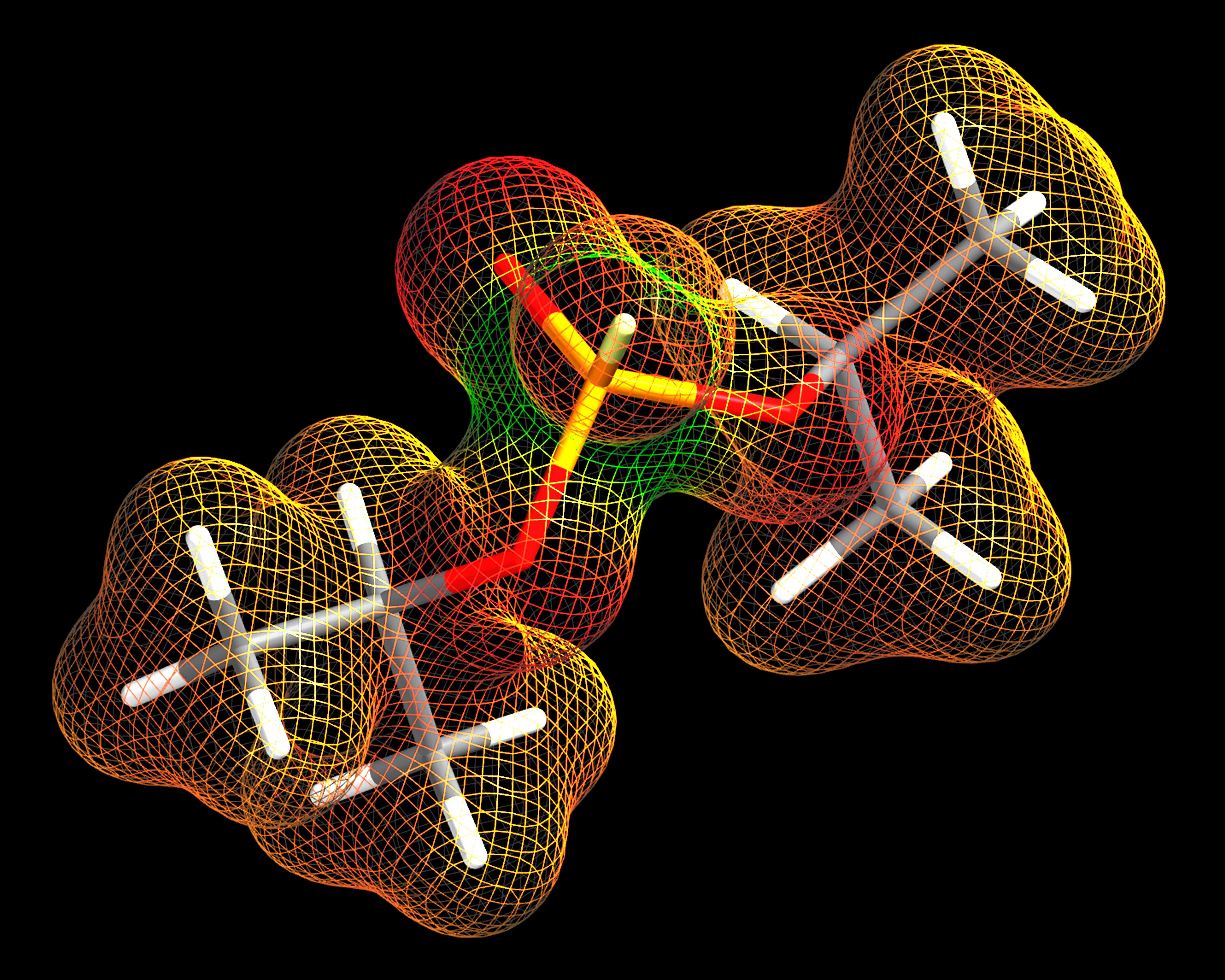
The bandage combines three key elements: a newly synthesized, brightly emitting red porphyrin; a green reference dye; and a wound sealant. The porphyrin and dye are phosphors, which absorb light and re-emit it at a longer wavelength, based on the level of oxygen present. At higher oxygen levels, which indicates healthy skin tissue, the reference dye shines green. At lower levels, which signals a more severe burn, the dye does not shine and the porphyrin gives off a reddish orange light. A doctor in the field can activate the phosphors using the flash from a smartphone camera, enabling accurate field diagnosis.
What’s more, the bandage continuously monitors the burn, and future versions will deliver therapeutics to the wound, according to Conor L. Evans, an associate professor at Harvard Medical School and researcher at Massachusetts General Hospital, who developed the bandage.
The road to recovery
Chan is medical director of a private plastic surgery practice, but he spends most of his clinical time tending military patients at burn center in San Antonio. There, burns to the face or hands are the most common injuries and may take many operations to repair before patients can perform simple tasks like brushing their teeth.
“Treating patients that are injured from war while serving their country allows me an opportunity to be able to help someone,” Chan says. “I feel like I’m doing something worthwhile.”
To explore careers in the U.S. Army Medical Department (AMEDD), click here.